X Ray Opacity Definition

Chest X Ray Shows A Well Defined Opacity In The Left Hilar Region Download Scientific Diagram

Chest Radiology

Chest X Ray Showed The Ill Defined Patchy Mass Opacity With Surrounding Download Scientific Diagram
Pa Chest X Ray Showing Ill Defined Opacity Right Lower Zone Arrow Download Scientific Diagram
Chest X Ray Showing Right Middle Lobe Opacity Arrow Download Scientific Diagram

Chest X Ray Fundamentals
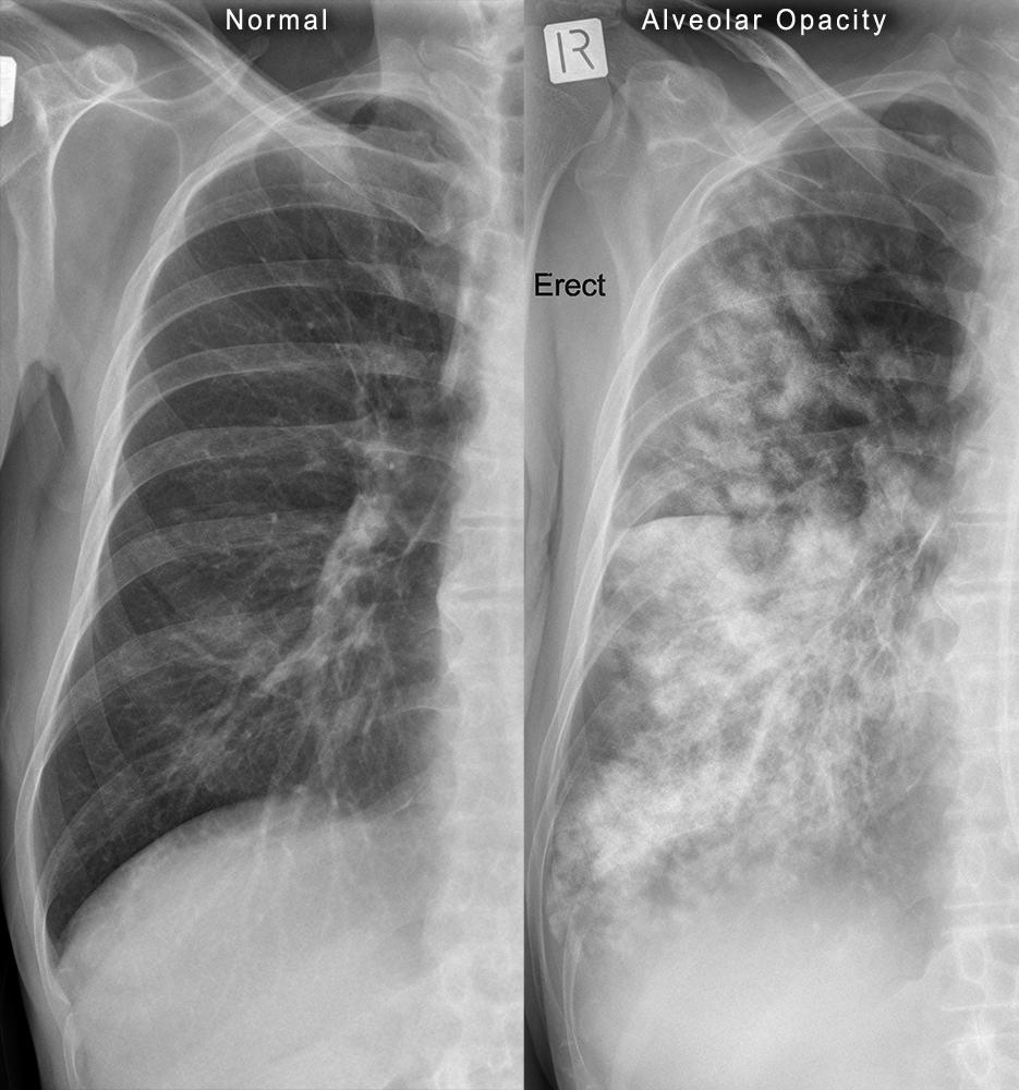
The key findings on the x ray are.
X ray opacity definition. In both of the cases above there is an abnormal opacity. A x e sup bt a component of opacity taking into account free acceleration mode. Pulmonary opacification represents the result of a decrease in the ratio of gas to soft tissue blood lung parenchyma and stroma in the lung. He is also the innovation lead for the australian centre for health innovation at alfred health and clinical adjunct associate professor at monash university.
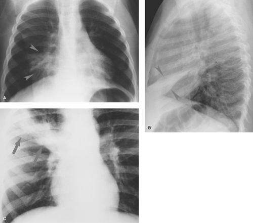
The basic diagnostic instance is to detect an abnormality. Sharply defined opacity obscuring vessels without air bronchogram. It is most useful to state the diagnostic findings as specifically as possible then try to put these together and construct a useful differential diagnosis using the clinical information to order it. Volume loss resulting in displacement of diafragm fissures hili or mediastinum.
He is a co founder of the australia and new zealand clinician educator network anzcen and is the lead for the anzcen clinician educator incubator programme. T time duration of free acceleration mode s. How to use opacity in a sentence. Chris is an intensivist and ecmo specialist at the alfred icu in melbourne.
It is one of the many patterns of lung opacification and is equivalent to the pathological diagnosis of pulmonary consolidation. A and b equation coefficients. In radiological studies it presents as increased attenuation of the lung parenchyma causing. Often used interchangeably with opacity density refers to an area on the x ray that is brighter than expected when x rays are absorbed or blocked by something such as the thick pus and mucous of a pneumonia this shows up as a brighter spot on the lungs.
Lobar atelectasis or lobar collaps is an important finding on a chest x ray and has a limited differential diagnosis. Where d sub 0 a measured value of opacity in a set mode at a maximum crank shaft speed.
Fundamental Radiological Findings Hazy Opacities Stepwards

Chest X Ray Showing Dense Homogenous Opacity In Right Middle And Lower Download Scientific Diagram

Multifocal Ill Defined Opacities Radiology Key

Chest X Ray Clearly Defined Homogenous Opacity With Pa Open I

X Ray Chest Showing Inhomogeneous Opacity In The Middle Zone Of Left Lung Download Scientific Diagram

This X Ray Shows The Ground Glass Opacity Composed By Diffuse Download Scientific Diagram

The X Ray Pns Revealed A Homogenous Opacity With A Convex Upper Margin Download Scientific Diagram

Chest X Ray Posteroanterior View Shows Ill Defined Patches Of Download Scientific Diagram

Chest X Ray Postero Anterior View Ill Defined Opacity Obscuring The Download Scientific Diagram

What Are Lung Opacities Kaggle

Normal Chest X Ray With Diaphragmatic Slips When The Lungs Are Hyperexpanded It Is Important Not To Confuse Blunting Of A Costophre Radiology X Ray Vision Eye

Chest X Ray Showing Non Homogenous Opacity In Left Upper Zone With Download Scientific Diagram
Pediatric Chest Radiology Key
