Contrast Definition X Ray

Kvp And Contrast Radiology Humor Radiology Radiology Imaging

Image Result For Ap X Ray View Mysuru X Ray Medical College

X Ray Imaging Can Be Used To Diagnose Whether A Patient Has Congestive Heart Failure Heart Failure Congestive Heart Failure Failure

Bronchopulmonary Aspiration Radiology Humor Radiology Radiology Technologist

Pin By Utheya Thevathas On Medical Radiation Sciences Radiology Imaging Radiology Schools Diagnostic Imaging

Radiographic Contrast Radiology Reference Article Radiopaedia Org
For example in an intraoral radiograph enamel will attenuate x rays more than dentin.
Contrast definition x ray. The term plain x rays is sometimes used to distinguish x rays used alone from x rays combined with other techniques eg ct. Contrast dye works by using substances that interfere with how the medical imaging equipment takes your images. Iodine circulatory system iodine has a particular advantage as a contrast agent because its innermost electron k shell binding energy is 33 2 kev similar to the average energy of x rays used in. For conventional radiography an x ray beam is generated and passed through a patient to a piece of film or a radiation detector producing an image.
Phase contrast x ray imaging pci or phase sensitive x ray imaging is a general term for different technical methods that use information concerning changes in the phase of an x ray beam that passes through an object in order to create its images. Radiocontrast agents used in x ray examinations can be grouped in positive iodinated agents barium sulfate and negative agents air carbon dioxide methylcelluose. For objects that attenuate more of the radiation than the adjacent tissue contrast is inversely related to object penetration. This test allows the radiologist to evaluate structures that are not clearly evident on conventional x ray exams.
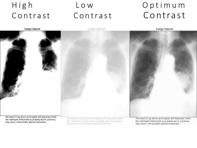
For example the contrast used in an x ray or ct exam is made of a substance that will block or limit radiation in certain parts of your body. Contrast is the difference in density or difference in the degree of grayness between areas of the radiographic image. X ray contrast is produced because x ray penetration through an object differs from the penetration through the adjacent background tissue. A comparison in which differences are demonstrated or enhanced.
This occurs because low energy radiation is more easily attenuated. Conventional radiography involves the use of x rays. This is a function of the number of x ray photons transmitted or the strength of the signals emitted by the two regions and the response of the recording medium. Therefore the ratio of photons that are transmitted through a thick and thin area will be greater with low energy radiation.
It refers to the difference in the intensity transmitted through the different parts of an object. 7 the radiographic contrast depends on the following three factors. This changes how the tissues that contain the medical imaging contrast appear on your images. Maximum 100 contrast is produced when no radiation penetrates the object.
In radiology the difference between the image densities of two areas is the contrast between them. A primary beam with greater kv results in an overall rise in penetration through all tissues decrease in attenuation differences therefore resulting in a lower contrast radiograph. Because bones block the x rays easily they show up clearly. Generating x rays using a low kilovoltage will generally result in a radiograph with high contrast.
Standard x ray imaging techniques like radiography or computed tomography ct rely on a decrease of the x ray beam s intensity attenuation when.

Pneumoretroperitoneum Is By Definition Presence Of Gas Within The Retroperitoneal Space On Plain Radiography The Differ Radiology X Ray Retroperitoneal Space

Abdomen Xray Shows Endovascular Coils Used To Treat Varicoceles In A Man With Infertility Radiology Radiologist Intervent Radiology Radiologist X Ray

Pin On Radtech

Chronic Calcific Pancreatitis With Pseudocyst Human Anatomy Human Organ Human
Ideal Radiography

The Radiology Assistant Heart Failure

Radiographic Quality Veterian Key

Lung Abscess A Single Contrast Enhanced Axial Ct Scan Image Of The Chest Shows A Large Cavitary Lesion In The Left Lower Radiology Imaging Radiology Ct Scan

The First X Ray X Ray Radiologic Technology Rad Tech Week

Bipartite Patella Radiology Case Radiopaedia Org Radiology X Ray Mri

Scattered Radiation And Contrast Radiology Schools Diagnostic Imaging Radiology
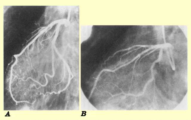
Image Noise

La Radiografia Lateral De La Columna Lumbar Revela Una Deformidad De Compresion En La Cuna Anterior Fractura Por Compresion Del Cuerpo Ver Painting Image Art
